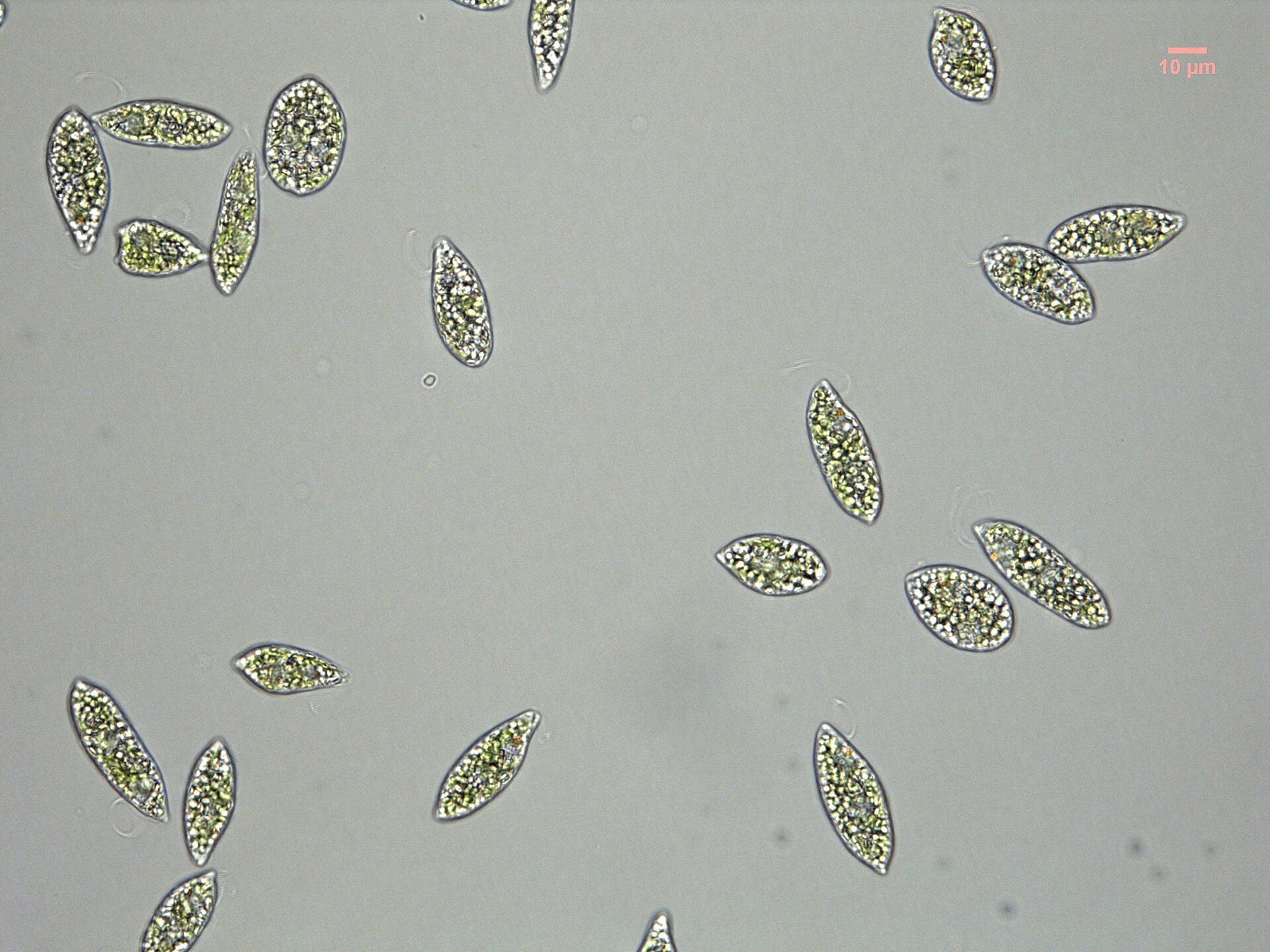
A single cell that steers by light with a red shade, a light sensor and a spinning body. It is not an animal, and its eyespot evolved separately from animal eyes.
FIG. A1.1Euglena gracilis cells under a light microscope. The small orange-red spot near one end of several cells is the stigma. Scale bar 10 µm.Photo: Ellis O'Neill, via Wikimedia Commons, CC BY-SA 3.0.
What the eye is
Euglena gracilis is a single-celled, photosynthetic flagellate, first described by Antonie van Leeuwenhoek in 1684.1 It belongs to the Euglenozoa, a group that also includes the parasites Trypanosoma and Leishmania.1 It is not an animal: the Euglenozoa sit in a separate branch of the tree of complex cells called Discoba.1 Its chloroplast came from a green alga that an ancestor took in and kept, an event called secondary endosymbiosis.12 It is a mixotroph, able to feed as well as photosynthesise.1 A cell is about 50 µm long and 5 µm wide.3
Beside the base of its single flagellum sits a red spot called the stigma, or eyespot.43 It lies in the cell’s cytoplasm rather than inside the chloroplast, as it does in green algae, and is made of globules of carotenoid pigment.45 Despite its name, the stigma does not detect light.4 The sensor is the paraflagellar body, a swelling at the base of the flagellum that the stigma partly shades.43
How it works
The paraflagellar body contains a blue-light-sensitive protein, photoactivated adenylyl cyclase (PAC), which turns light directly into a chemical signal inside the cell.6 When Iseki and colleagues (2002) blocked the production of PAC, cells stopped turning away from a sudden rise in light.6 Ntefidou and colleagues (2003) found that blocking it also stopped cells steering towards or away from light.7
A single shaded sensor gathers information about direction only over time, as the body carries it round.8Euglena swims at about 50–100 µm a second while rolling about its long axis 1–2 times a second, beating its flagellum 15–20 times per roll.3 As it rolls, the stigma passes between the light and the sensor, so the signal switches on and off once per turn.3 That signal changes how the flagellum beats, and the cell turns.3 Land and Nilsson (2012) compare this to an animal swinging a single shielded detector from side to side to find which way is brighter.8
FIG. A1.2The stigma sits beside the light sensor at the base of the flagellum. As the cell rolls, the stigma shades the sensor once per turn, and the change in signal steers the cell.Drawing: Half an Eye, CC BY 4.0. After Tsang et al. 2018, fig. 1d, and Jékely 2009.
Euglena can steer towards light or away from it, depending on conditions.73 When the light jumps suddenly, cells swim in polygons of straight runs and sharp turns, or spin on the spot in very bright light; switching between these lets them back away from bright patches and move down a light gradient.3 The stigma is needed for this: mutants without one lose normal phototaxis and swim in two preferred directions instead of one.4
What it tells us about evolution
Steering by light has evolved at least eight times among eukaryotes, the organisms whose cells have a nucleus.4 Different groups do it with different light-sensing proteins: green algae use one kind of rhodopsin, euglenids use PAC, and animals use their own opsins.4 The opsins animals see with are not thought to share an origin with those of green algae.8
FIG. A1.3Euglenids and animals sit on separate branches of the tree of complex cells. Steering by light evolved several times, and each group uses its own light-sensing protein.Drawing: Half an Eye, CC BY 4.0. After Burki et al. 2020, fig. 1, and Jékely 2009, table 1.
The method, though, is shared: a sensor with a restricted view, a shade on one side, and a body that spins as it swims.4 The Euglena eyespot is therefore not an ancestor of animal eyes. It is a separate solution to the same task, and it shows how little that task needs: one shaded sensor and a rotating body.84
One research group has argued that the paraflagellar body is a crystal of a rhodopsin-like protein.9 A 2017 review by Häder and Iseki describes a long debate over the receptor and treats PAC as the one identified.10