4.1 A series of useful eyes
In On the Origin of Species, Charles Darwin argued that if a graded series of eyes could be shown to exist, from simple to complex and each useful to its owner, the difficulty of believing that the eye evolved by natural selection “can hardly be considered real”.12 He noted that the ideal series would come from an organ’s direct ancestors, but since this is “scarcely ever possible”, we have to look instead at living species of the same group.1 This chapter follows that series through living animals, one step at a time, and then looks at a calculation of how long the whole journey might take.
Chapter 1 sorted light-guided behaviour into four classes: non-directional light monitoring, directional photoreception, low-resolution spatial vision and high-resolution spatial vision.345 Each class matches a stage of eye structure: unshielded photoreceptors, pigmented ocelli (small, simple eyes), pigment pits or cups, and eyes with focusing optics.6 Nilsson (2009) links each new class to a key innovation: efficient light-sensitive pigments, screening pigment that lets a receptor tell direction, folded and stacked photoreceptor membrane, and finally focusing optics.47
The task sets the minimum the eye must do. Directional photoreception needs only a screen that shades the receptor on one side; for phototaxis, swimming towards or away from light, a field of view as wide as 180° is enough, because the animal finds the direction by scanning as it moves its body.8 Many low-resolution tasks need a resolution of only about 5–30°, and pigment pits and cups can support those tasks, though not high-resolution ones.6 Spotting prey and predators needs resolution no worse than a few degrees, which requires a lens or other focusing optics; for comparison, humans resolve about 1/60 of a degree.6 Land and Nilsson suggest that improved sensitivity, more than resolution, was probably the main reason lenses evolved, a hypothesis section 4.6 returns to.69
Land and Nilsson also argue that most eyes, simple or advanced, are probably close to optimal for what their owners need them to do.10 On that view, the series runs from simple tasks done well to complex tasks done well.10
4.2 Sensing light without an eye
Some light-guided organisms manage without a nervous system. The larva of a box jellyfish carries about 20 ocelli, each a single cell that forms its own pigment cup, holds light-sensitive microvilli (finger-like folds of membrane) and bears a cilium that steers the larva.1112 The larva has no nervous system at all: each cell senses light and drives the steering itself.11
Euglena gracilis is a single-celled alga with a pigmented eyespot near the base of its flagellum, the whip-like tail it swims with, and it swims towards light.13 The pigmented spot is not the light sensor; it works as a shade.1415 Molecular studies point instead to a blue-light-sensitive protein, photoactivated adenylyl cyclase, which in normal cells sits in a structure called the paraflagellar body.1415 This protein is the receptor for the step-up photophobic response, the cell’s reaction to a sudden rise in light, and for steering both towards and away from light.15 When its production is knocked down, the cell’s steering by light is suppressed.1415
Some dinoflagellates, another group of single-celled organisms, go much further, with a lens and a retina-like structure inside one cell.16 They evolved this on their own, and it has little bearing on how animal eyes evolved.16
4.3 The two-cell eye
The simplest animal eyes are eyespots of just two cells: a photoreceptor and a pigment cell that shades it.1712 Jékely and colleagues (2008) noted that such eyespots resemble Darwin’s “proto-eyes”.17
They studied the larva of the ragworm Platynereis, which swims with bands of beating cilia, the hair-like structures on its surface.17 Light falling on one eyespot changes the beat of the nearby cilia through a direct nerve connection that uses the transmitter acetylcholine, and this steers the larva.17 The larva also spins about its long axis as it swims, and this rotation is essential for phototaxis; the helical path it produces makes the steering more precise.17
As the larva grows, it adds four adult eyes, each with only 2–7 photoreceptors, a few pigment cells and a lens.18 Randel and colleagues (2014) mapped every neuron and connection behind these eyes: a circuit of 71 neurons joined by 1,106 synapses.18 The circuit compares the light reaching the two sides of the body and bends the body when the two differ.18 Three- and four-day-old larvae show mixed phototaxis, some swimming towards light and others away.18 When the eyes on one side were lit, larvae bent towards or away from that side, and some switched between the two on repeated stimulation.18 The authors suggest that phototaxis of this kind may have been the first visual task animals performed.18
4.4 The cup
Once a receptor is shaded, spatial vision can grow in two ways: by placing more receptors under one shield, which leads to single-chambered eyes, or by repeating the whole shaded unit many times, which leads to compound eyes.19 This chapter follows the first route; compound eyes have a chapter of their own.
Spatial information comes from comparing receptors that view different directions.2021 In a pigment pit, each receptor’s view is limited only by the shadow of the pigment behind it, so which receptors are lit shows where the light is coming from.2021
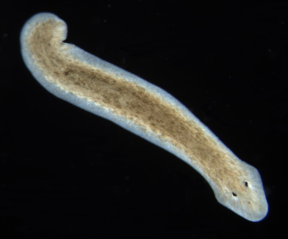
A planarian flatworm’s eye is a cup formed by a single pigment cell and lined with photoreceptors whose axons, the cells’ output fibres, point towards the light.12 Its resolution is about 35°, the coarsest of all the eyes in Land and Nilsson’s table of eye performance.22
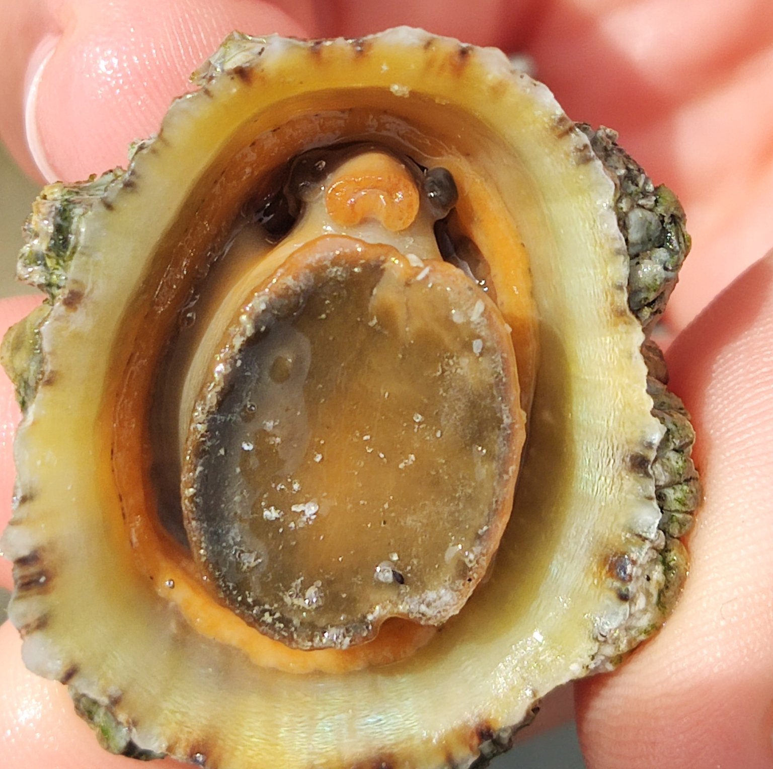
Molluscs show the same stage. The limpet Patella has a V-shaped pigmented pit, and each of its receptors accepts light from a range of 90° or more.21 That is enough for the limpet to find lighter or darker areas.21


4.5 The pinhole
In the abalone Haliotis the mouth of the pit is drawn inwards, leaving a narrower opening, and each receptor’s view shrinks to perhaps 10°.23 Narrowing the opening further gives a sharper image but lets in less light, and Nautilus is the only animal that has taken this route to its end.24
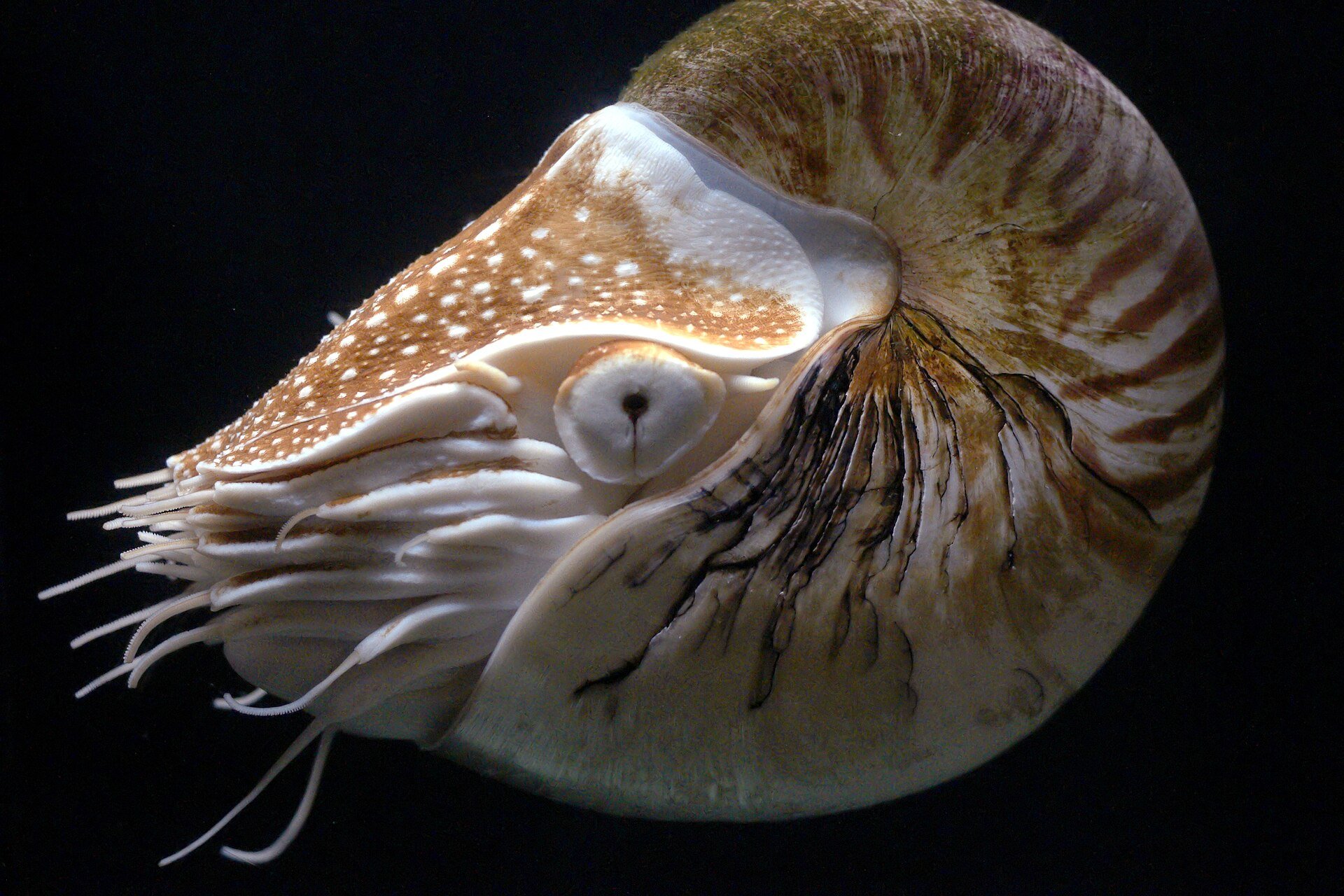
The eye of the chambered nautilus, Nautilus pompilius, is about 1 cm across, has no lens or cornea, and is open to the seawater.242526 Its pupil widens and narrows with the light level; Land and Nilsson give a range of 0.4 to 2.8 mm, while Schwab reports at least 0.7 to 2.25 mm.2526 Muscles rotate the eye to keep its vertical axis steady as the animal rocks.25
Muntz and Raj (1984) tested the nautilus with optomotor experiments, which measure whether an animal turns to follow a moving striped pattern, and put the finest separation it can detect at roughly 6–11°.27 Stopping the pupil down to 0.4 mm would narrow each receptor’s view to about 2.3°, but the image would then be about 400 times dimmer than in a fish eye.28 This is the cost of a pinhole: sharpness is bought with light.2428
Whether the nautilus eye is a survivor of an ancient pinhole stage is uncertain. Fossils are usually read as showing that its lensless eye is ancestral.29 Two genetic studies suggest otherwise.3029 The gene six3/6, and crystallin genes downstream of it, are not expressed in the developing nautilus eye.30 The nautilus genome also shows gene losses and a reduced family of crystallin genes, which its authors read as plausible signs of degeneration from a more complex organ.29 The nautilus is therefore best treated as a working example of a pinhole eye, not as proof of an ancestral stage.
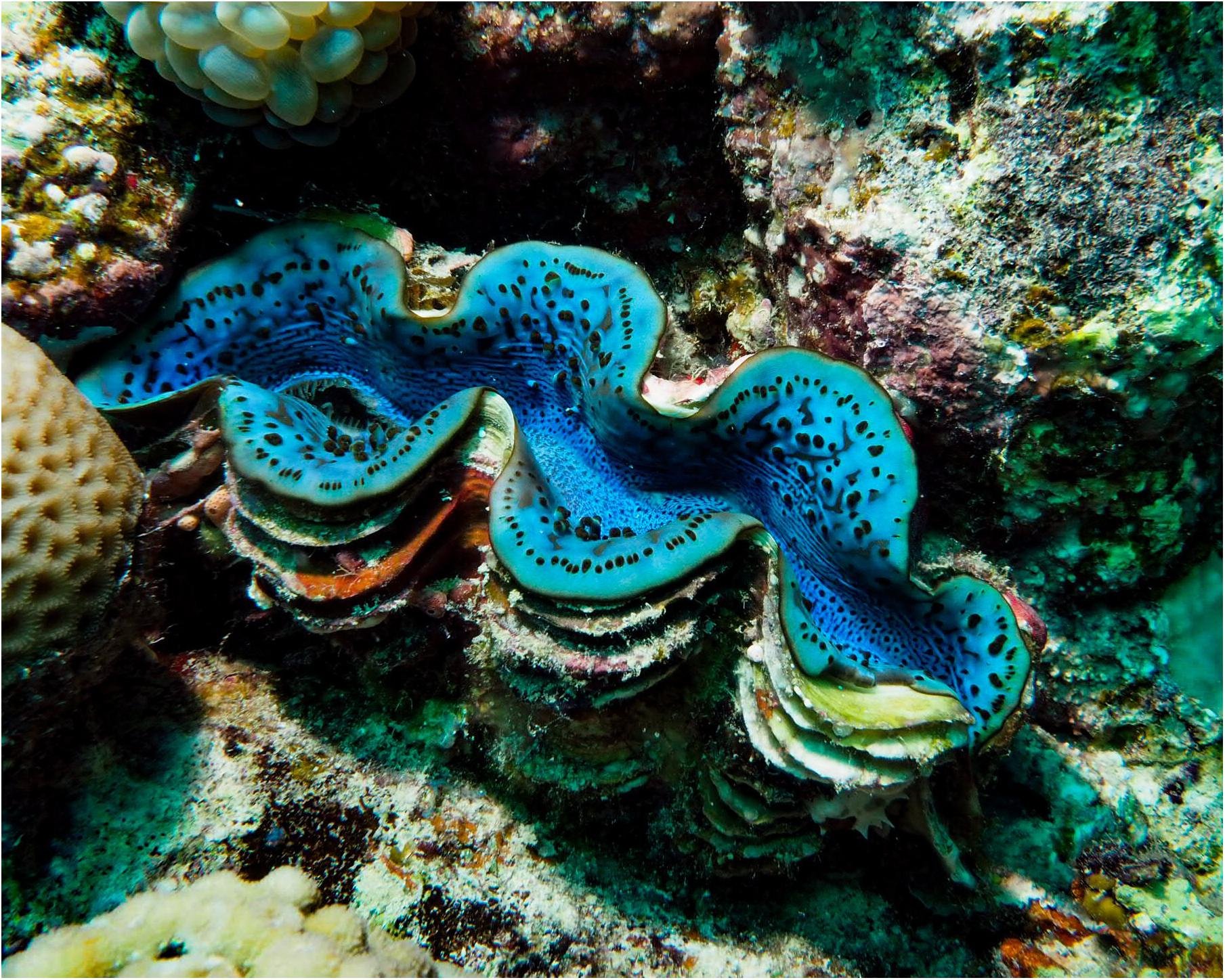
Giant clams, Tridacna, also have pinhole eyes, up to a thousand of them around the edge of the mantle, the soft tissue exposed between the two halves of the shell.25 Each eye is a pit about 0.5 mm wide and deep with an opening of 0.1 mm, holding about 250 receptors; in Tridacna maxima each receptor views an angle of about 16.5°.25 A clam withdraws its siphons and closes its mantle when a shadow passes over it.25 It also reacts to an object that casts no shadow, if the object fills an angle comparable to or larger than one receptor’s view, so a 10 cm fish triggers a response at about 42 cm.25 Land and Nilsson suggest that clams have not evolved sharper eyes because 16.5° is good enough, and that finer vision would bring more false alarms from harmless animals and debris.25


4.6 The lens
A lens solves the pinhole’s trade-off by bending light, so that a wider opening can give the same sharpness.9 Snails show what simple lenses can look like. In the land snail Helix the lens is a ball of jelly that converges light a little without forming a sharp image.24 In the periwinkle Littorina the lens has a graded refractive index, meaning that how strongly it bends light changes from centre to edge, and it forms a good image.24
Eyes like the snail’s, in which a lens-like body fills the cup without bringing light to a sharp focus on the retina, are called under-focused lens eyes.31 They are common in gastropods and polychaete worms, and sit between cup eyes and camera eyes.31
Box jellyfish carry 24 eyes of four types.32 They sit on four sensory structures called rhopalia, each carrying two lens eyes and two pairs of pit eyes.3133 A heavy crystal keeps each rhopalium hanging so that its small upper lens eye always points straight up and its larger lower lens eye points obliquely down and inwards, whatever the jellyfish’s orientation.3133 Nilsson and colleagues (2005) found that the lenses in these eyes have a finely tuned gradient of refractive index that gives a nearly aberration-free image, yet the retina sits too close to the lens for that image to be in focus.3234 Even so, the lens pays its way: without one, an eye with the same degree of blur would need an opening slightly under half the width and would catch about one-fifth as much light.9
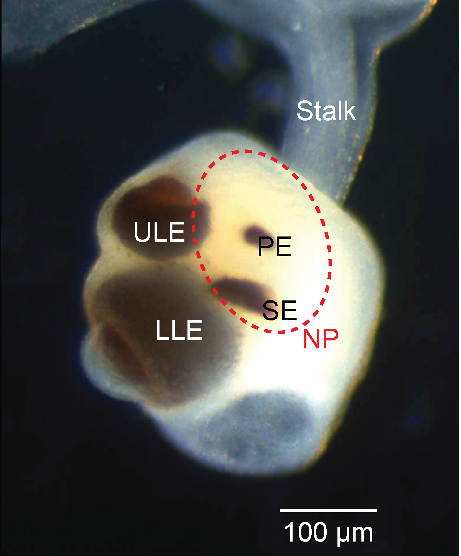

The lens eyes serve a few tasks.31 In the box jellyfish Tripedalia cystophora, the lower lens eyes guide the animal around obstacles such as mangrove roots.3531 In flow-chamber tests, avoidance was guided by vision: it was triggered when an obstacle filled a certain angle of the visual field, and tended to follow how strongly the obstacle contrasted with its surroundings.35 The upper lens eyes look up through the water surface at the mangrove canopy, and the jellyfish use this view to find their way back to their preferred habitat at the edge of mangrove lagoons.3331 The photoreceptors of the lens eyes are slow, work over a range of about 3 log units of light intensity (a thousandfold) and contain a single opsin, the light-sensitive protein of animal photoreceptors, so the jellyfish are almost certainly colour-blind.36
A related species, Chiropsella bronzie, has lens eyes that are even further out of focus: in its lower lens eye the blur spreads each point of light over 20° with the pupil closed and 52° with it open, and the lens of its upper eye has no significant optical power.3738 Land and Nilsson argue that the blur is useful: it passes the coarse image detail these behaviours need and removes fine detail that would not help, and small changes in the retina’s position or the lens’s gradient could tune each eye to its task.9
Why lenses first appeared is still debated, and the two leading ideas come from the same research group. One is that lenses first raised sensitivity, letting a cup eye gather more light.69 The other is that they first tailored each eye’s view to a particular task.32 The two are not exclusive, and both remain hypotheses.
Land and Nilsson also suggest that material may first have filled eye cups for reasons unrelated to focusing, such as screening out short-wavelength light or supporting the retina, and only later became dense enough to focus.39 The nautilus eye cup holds no such material, which they suggest may be why it never evolved a lens.9
4.7 Building a better lens
A lens made of protein runs into a problem of geometry. A uniform sphere of dry lens protein in seawater would have a focal length of about 4 lens radii, but the lenses of fish and cephalopods focus at about 2.5 radii, a figure known as Matthiessen’s ratio.40 Real lenses reach it because their refractive index falls from the centre to the edge, which both shortens the focal length and corrects spherical aberration, the blur that arises when rays through the edge of a lens focus at a different distance from rays through its centre.41 A central index of 1.52 is enough to reach the 2.5 ratio.41
Snails show the range of focused lens eyes that results. The freshwater snail Lymnaea has an eye only 100–200 μm across, with an excellent lens and a fovea-like pit in its retina.42 At the other extreme, the predatory sea snail Pterotrachea has a 3 mm eye with a large spherical lens and a retina only six rows of cells wide.42
Lenses of this kind have evolved separately in fish, in cephalopods, in gastropods (perhaps more than once), in alciopid polychaete worms and in one copepod, a tiny crustacean called Labidocera.43 More broadly, lens eyes evolved from pigmented pit eyes independently in at least four phyla.44 Chapter 7 counts these separate origins.
The proteins that make up a lens are called crystallins.45 Most were recruited from proteins that already had other jobs, mainly enzymes and heat-shock proteins, which help cells cope with stress; what changed was where and how much of them the cell made.114546 Some crystallins are the very same gene product as an enzyme: ε-crystallin, for example, is lactate dehydrogenase B.47
4.8 How long would it take?
In 1994 Nilsson and Pelger estimated how many generations natural selection would need to turn a flat patch of light-sensitive cells into a camera-type eye with a focusing lens.48 Their paper is an analytical calculation using formulae from population genetics; it contains no computer simulation.48
The model starts with a flat patch of light-sensitive cells lying between a transparent protective layer and a layer of dark pigment.49 Selection acts on one property only, spatial resolution, and the overall width of the organ is held constant throughout.49 At first, the fastest way to improve resolution is to deepen the patch into a pit, until the pit is as deep as it is wide.49 After that, narrowing the opening improves resolution faster, and the eye becomes more like a pinhole.49
The paper sets out the sequence as eight stages (Figure 4.9). In stages 2 and 3 the photoreceptor and pigment layers, together called the retina, fold inwards into a hemisphere, while the transparent layer thickens into a vitreous body.50 The vitreous body’s refractive index is 1.35, too low to have any significant optical effect.50 In stages 4 and 5 the retina keeps growing without changing its curvature, so the change shifts from deepening the pit to narrowing its opening.50 The authors chose the aperture of stage 6 to match the proportions of real eyes of this type.50
Narrowing has a limit set by photon noise, the random variation in how many particles of light arrive.50 There is an optimum aperture, beyond which resolution cannot improve without a lens.50 In the model, even the weakest lens is better than none.51 In stages 6 to 8 the lens first appears as an ellipsoid filling the aperture, then becomes spherical and moves to the centre of the retina’s curvature.51 Meanwhile the old aperture stretches into a flat iris, and the focal length shortens from three times the distance between lens and retina to twice that distance and then to equal it, so the image is sharp only at stage 8.51 The lens appears as a local rise in refractive index, from 1.35 to 1.52, and the sequence ends with a focal length of 2.55 lens radii, close to Matthiessen’s ratio.51
Throughout, the model widens the receptors so that each catches the same number of photons, which keeps the ratio of signal to noise constant.51 Relative to stage 1, receptor diameter grows to 1.95 at stage 3 and 4.56 at stage 5, and stays between about 4 and 5 after that.51
Stage 8 of 8
- Receptor diameter, d
- 4.1
- Lens focal length, f
- P
- Steps of 1%
- 1,829 in all
- Receptor layer
- Pigment layer
- Protective layer, then vitreous body
- Lens
d is receptor diameter relative to stage 1; P is the distance from lens to retina.
Nilsson and Pelger then counted how many steps of 1% change the whole sequence needs: 1,829.51 The steps divide between the seven transitions from one stage to the next as 176, 362, 270, 225, 192, 308 and 296, and resolution improves roughly evenly throughout, with no stretch in which change brings no gain.51 They measured each structure’s change on its own, as if no change followed passively from any other.46 Doubling a length takes 70 steps of 1%, so 1,829 steps amount to a change of about 80 million times, as much as lengthening a 10 cm finger to 8,000 km.46 The authors acknowledge “unavoidable ambiguities” in how change in shape can be measured.46
4.9 From steps to generations
To turn steps into generations, the authors used a formula in which the change per generation depends on heritability (how much of the variation in a trait is passed on), the intensity of selection and the amount of variation in the population.52 They took heritability as 0.50 and deliberately set selection intensity and variation low, at 0.01 each, which gives a change of 0.005% per generation and a total of 363,992 generations.52 They also assumed that every step happened one after another, never in parallel, to keep the estimate pessimistic.46
The table divides that total between the stages of the model.5152
| Transition | Steps of 1% | Generations (about) |
|---|---|---|
| Stage 1 to 2 | 176 | 35,000 |
| Stage 2 to 3 | 362 | 72,000 |
| Stage 3 to 4 | 270 | 54,000 |
| Stage 4 to 5 | 225 | 45,000 |
| Stage 5 to 6 | 192 | 38,000 |
| Stage 6 to 7 | 308 | 61,000 |
| Stage 7 to 8 | 296 | 59,000 |
| Total | 1,829 | 363,992 |
Generations per transition are our arithmetic from the paper’s step counts and its rate of 0.005% per generation; the total is the paper’s own.
At one generation a year, a camera-type eye could evolve in under 364,000 years.53 The authors note that, on this estimate, eyes could have evolved more than 1,500 times since the early Cambrian.53
Over the same sequence, the eye’s performance climbs steadily. Measured as the number of image points the eye can resolve across its hemispherical field of view, it rises from about 1 at stage 1 to about 70 at stage 7, in an almost straight line on a logarithmic scale.46 Stage 8 is left off the paper’s graph because its performance depends on light level and eye size, and could be up to six orders of magnitude, a millionfold, higher than stage 7.46
How cautious were the inputs? Across 1,120 heritability estimates from wild animal populations, morphological traits, those of body form and size, averaged 0.46.54 The paper’s heritability of 0.50 is therefore close to typical.5254 Its selection intensity was not. Reviews of more than 2,500 estimates from 63 field studies of 62 species found that the strength of directional selection on a trait, measured in standard deviations of that trait, has a median of 0.16, with few estimates above 0.50.5556 The paper used 0.01.52
With typical measured values in place of the paper’s (heritability 0.46 and selection 0.16, keeping its variation of 0.01), the same formula gives a far shorter time: about 0.074% change per generation, roughly 15 times the paper’s rate, or about 25,000 generations in place of 363,992.525455 This is arithmetic, not an estimate of how long eyes took to evolve, for three reasons. First, the field figure is a selection gradient, which matches the paper’s selection intensity only when no correlated traits are also under selection, though Kingsolver and colleagues found such indirect selection was usually modest.55 Second, field estimates are probably inflated, because most studies lacked the statistical power to detect the selection they reported and publication bias may push published values upwards.57 Third, one review found that the direction of selection often changes from year to year, while a later reanalysis that corrected for sampling error found it largely consistent; neither covers spans anywhere near 364,000 generations.5859 What the comparison does show is that the 1994 inputs for selection were cautious.525556
The model has stated limits. It starts with photoreceptors already in place and covers the optical geometry of the eye only.2052 Nilsson and Pelger treat the iris, focusing adjustment (accommodation), the blood-carrying choroid, the sclera and neural processing as refinements that the sequence does not need.2052 The origin of photoreceptors is the subject of Chapter 2.
4.10 Why the intermediate stages survive
Eyes resembling the stages of the model exist in living animals.5260 Nilsson and Pelger point out that molluscs and annelid worms alone show the complete series.52 Across the animal kingdom, gastropods have pigment-cup eyes, under-focused lens eyes and aquatic camera eyes; cephalopods have pinhole-type cup eyes and camera eyes; and polychaete worms have cup eyes, intermediate eyes, camera eyes and compound eyes.61
Intermediate eyes persist because an eye is only worth what its owner can do with it.5319 A sluggish worm, Nilsson and Pelger point out, would gain nothing from a fish’s eyes without a fish’s brain and body to use them.53
Key evidence
- Light-guided tasks fall into four classes, and each class matches a stage of eye structure, from a shaded receptor to a focusing lens (4.1).
- Working eyes exist at every stage: single-cell ocelli, two-cell eyespots, pigment cups, pinholes, under-focused lenses and graded-index lenses (4.2–4.6).
- Even out-of-focus lens eyes do useful work: box jellyfish use theirs to avoid obstacles and to find their way home (4.6).
- A deliberately pessimistic calculation puts the change from flat patch to camera-type eye at 1,829 steps of 1%, or about 364,000 generations, and field measurements show its selection input was cautious (4.8, 4.9).
- Intermediate eyes survive because each suits its owner’s way of life (4.10).